
DENTISTRY
Black Diamond Equine offers complete dentistry care for your horse. Equine dentistry is a key part of comprehensive equine health care, which includes an evaluation of a horse’s entire mouth (teeth, gums, jaw and tongue) for signs of disease as well as a horse’s entire body. Equine teeth continuously erupt throughout the life of the horse and the development of sharp points and hooks can occur over time based on how they chew and break down feed material. Because of this, horses require annual dental attention to prevent the development of permanent dental disorders, irritation to soft-tissue structures in the mouth, dropping feed, and weight loss. The inability to breakdown plant material can lead to impaction of the intestinal system and development of signs related to colic.
Common signs of dental problems include:
• Dropping feed or small bundles of chewed hay
• Difficulty chewing or excessive drooling
• Bad breath
• Poor performance, reluctance to exercise
• Fighting or chewing the bit, head tossing
The Oral Examination:
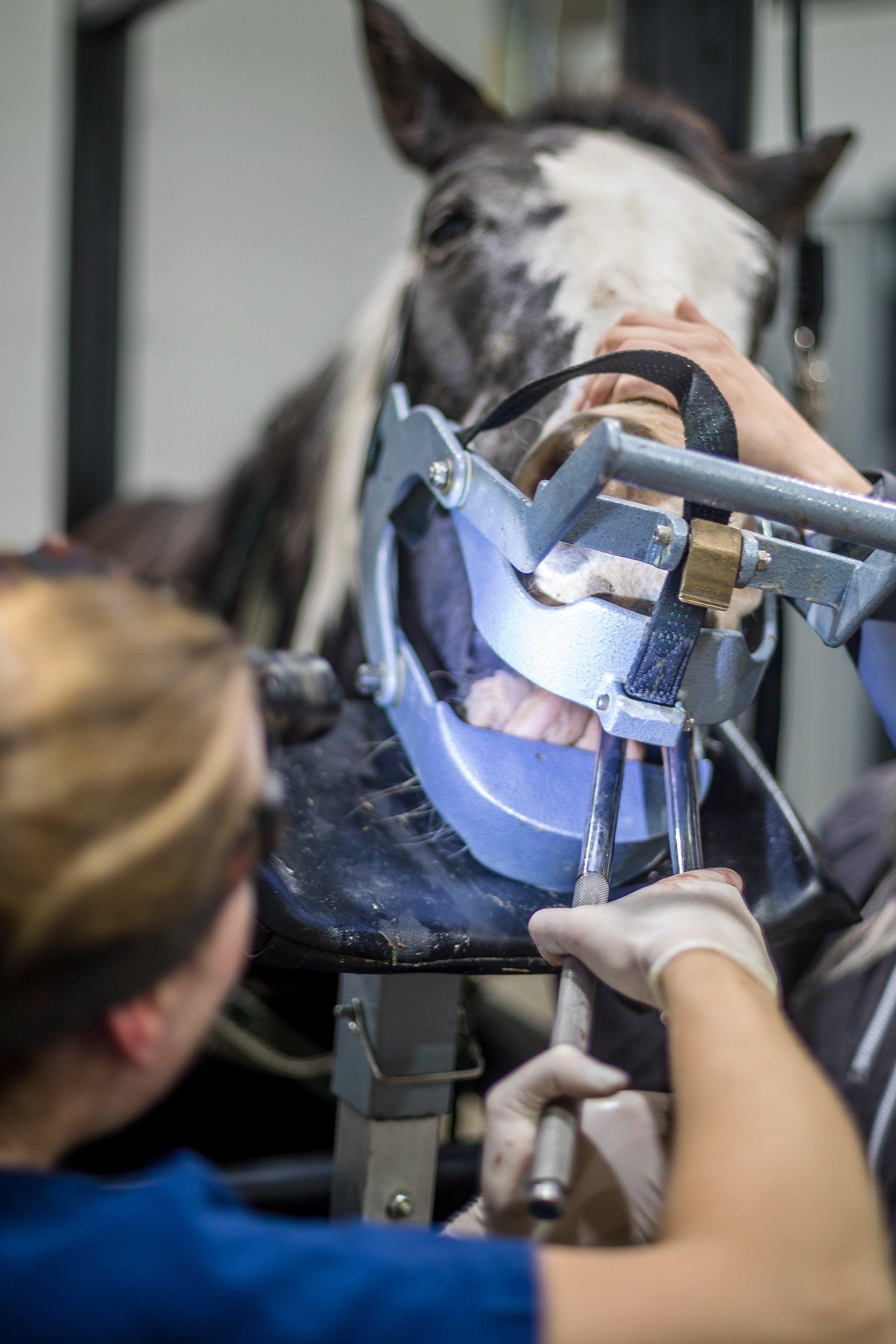
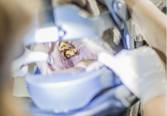
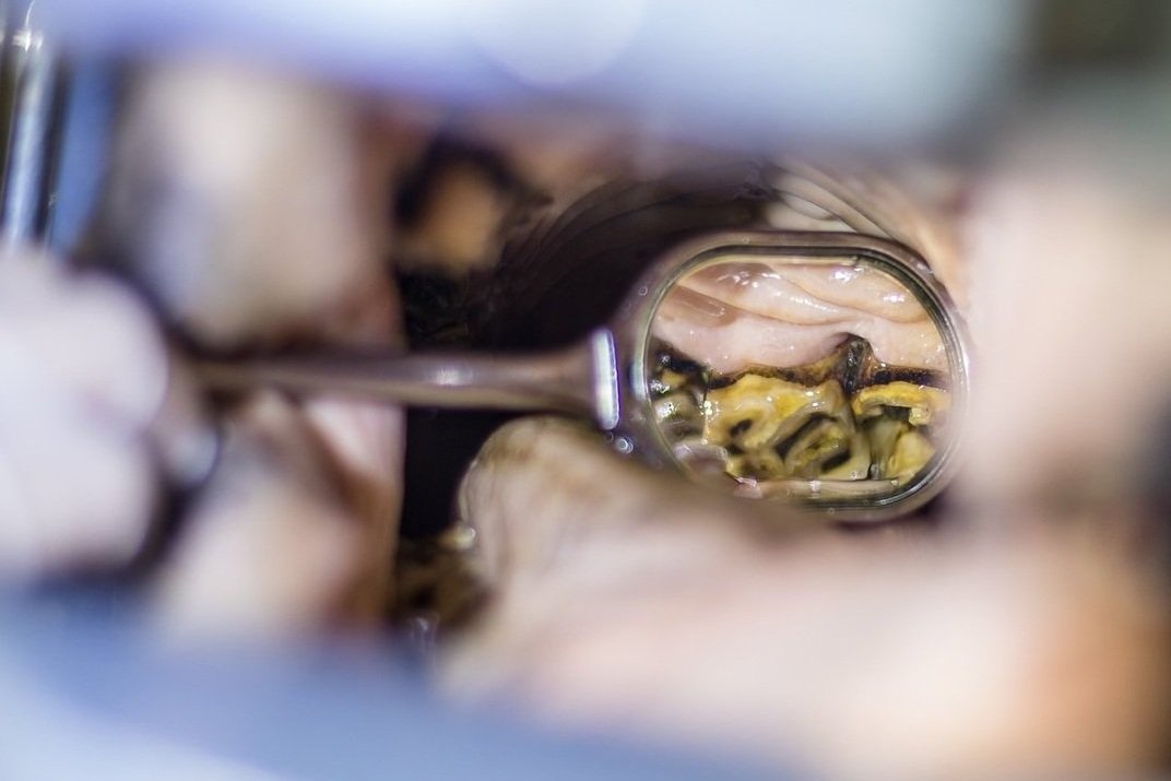
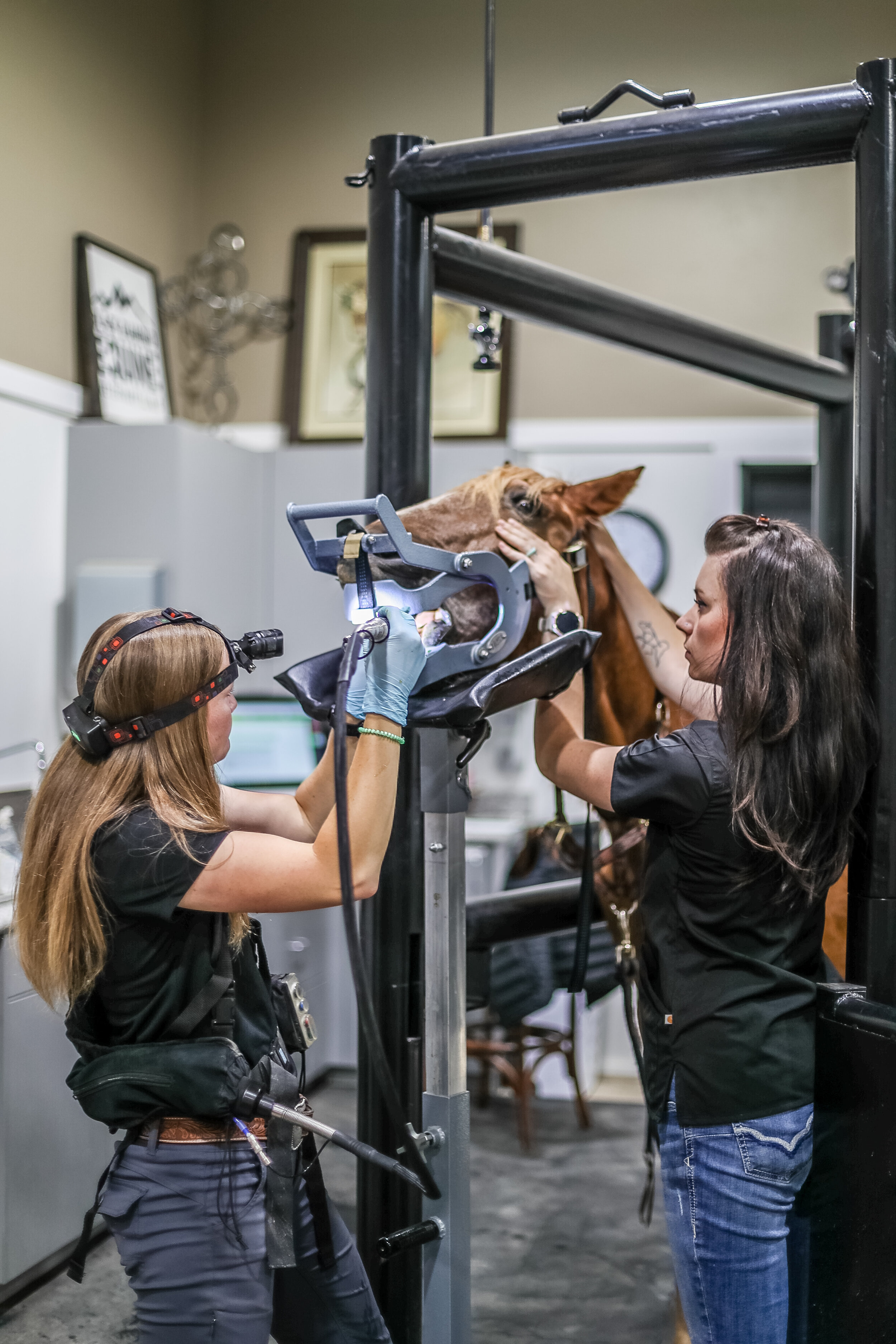
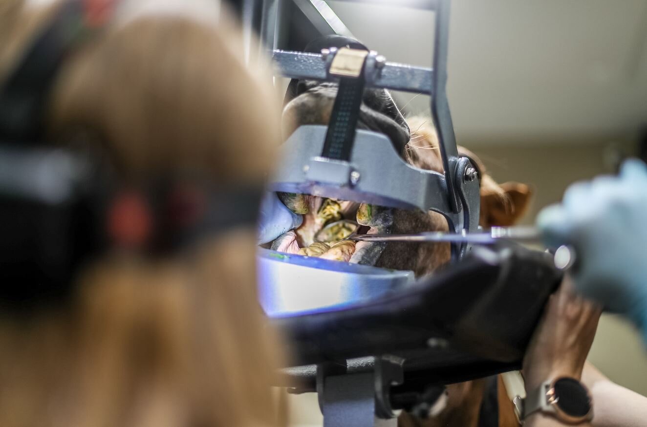
A complete oral examination looks at more than just the teeth. A thorough examination of the oral soft tissues, occlusal surface of the teeth and periodontal health around the roots of each tooth requires a sedated examination with a dental speculum in place. A comprehensive examination involves utilizing a mirror or oral endoscope to visualize hard to see places in the mouth.
The occlusal adjustment “Float”:
The term “Floating” a horse’s teeth was derived from a practice in masonry where a file was used to level bricks. Over the years, equine dentistry has expanded beyond simply leveling the teeth within the mouth and a focus has been adjusted towards returning balance within the mouth to prolong the life of each tooth and promote overall good oral health.
Sharp enamel points: These are points that form on the cingulae of the molars and can cause significant ulceration of the cheek pouches if left unaddressed.
Utilizing water cooled dental instruments reduces thermal damage to the teeth while adjustments are being performed and therefore reduces the likelihood of pulp damage during corrective dentistry.
Maloclusions:
Abnormalities in tooth eruption, chewing patterns, periodontal disease and jaw conformation can predispose horses to developing uneven chewing surfaces that can significantly impair their ability to process feed. If identified early, these abnormalities can be managed with routine occlusal adjustments. Examples of abnormalities encountered include:
Hooks
Steps
Wave mouth
Shear mouth
Excessive transverse ridges


Periodontal disease management
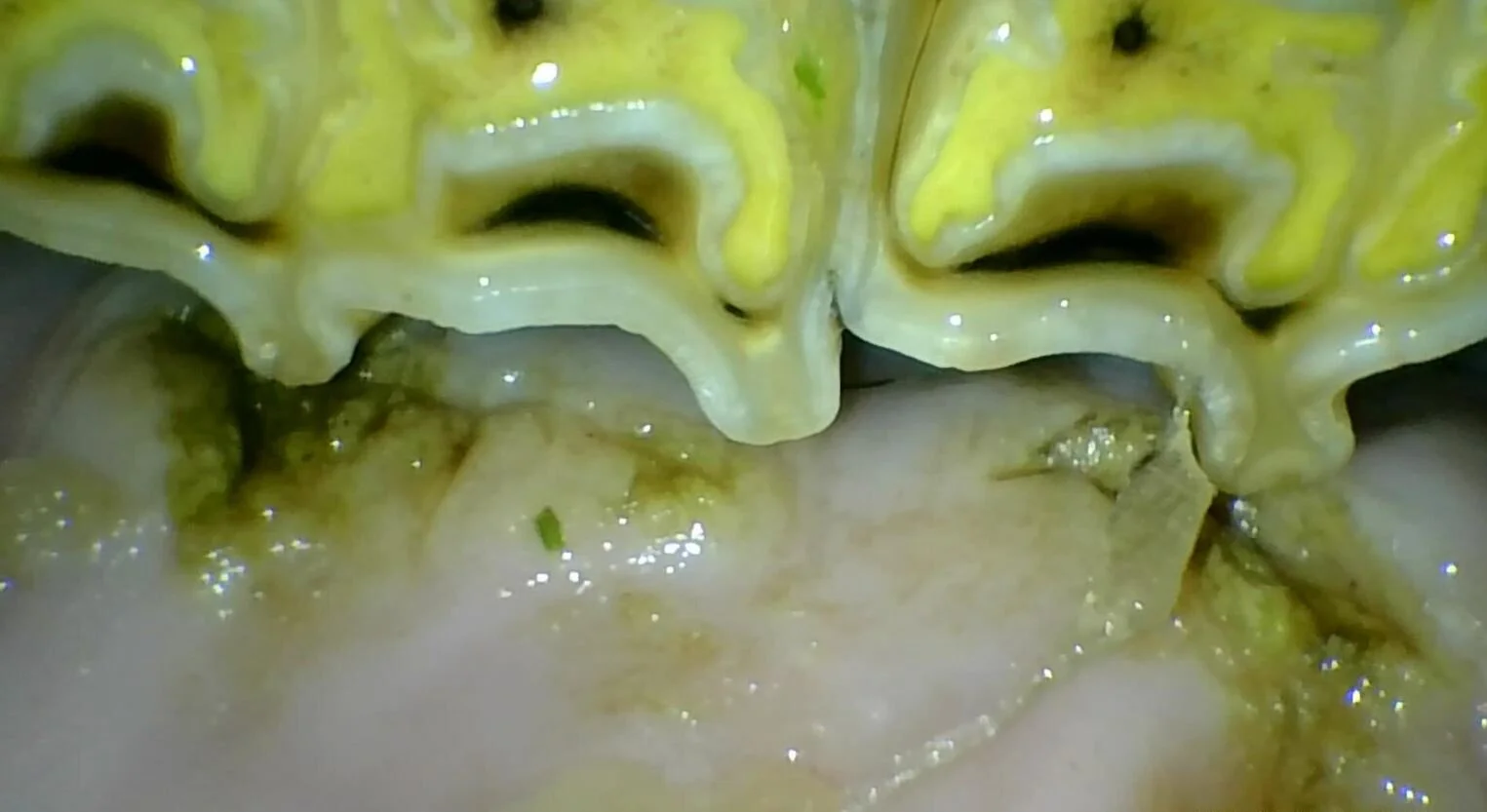
Periodontal disease is a common finding on a thorough examination of the horse’s mouth. Periodontal disease frequently is seen when feed along the gumline or between teeth accumulates which causes bacterial overgrowth, gingival inflammation and degradation of the clinical crown of the tooth. Horses may develop “cavities” that can have serious consequences for the health of the teeth and surrounding structures. Diagnosis of periodontal disease and implementation of appropriate treatment procedures is important to prolong the health of the tooth and ultimately, of your horse.
Diagnostic imaging:
Digital Radiographs: Advancements in digital radiography have improved our ability to detect dental disease early on and achieve high quality images to help determine what structures are affected. These images allow us to evaluate the sinus cavities, individual tooth roots as well as the adjacent bone supporting these teeth.
Oral Endoscopy: This is a powerful imaging modality in which a small camera is inserted into the mouth while a speculum is in place. The magnification of each individual tooth and surrounding soft tissues allows for an incredibly thorough and detailed examination. These images are useful in performing delicate procedures or enhancing visualization in the farthest corners of the mouth
Sinoscopy: The teeth are intimately associated with the sinus cavities of the horse’s skull. Endoscopic evaluation of the sinus cavity can be used to obtain the most accurate diagnosis with complicated sinus conditions and plan procedures accordingly.


Extractions:
Intraoral Extraction of cheek teeth, incisors and canines is performed under standing sedation. Local blocks are performed to numb the surgical site and allow for patient comfort while the procedure is completed. Teeth may need to be extracted due to advanced periodontal disease, crown fractures, pulp horn exposures or disease of the roots.
Partial Coronectomy of a tooth may be used to aid complicated extractions by removing a small section of the crown of the tooth with a high speed drill in order to improve the amount of space available to loosen the tooth and successfully achieve intraoral extraction.
Sectioning of the tooth may be required if the tooth is not amenable to traditional intraoral extraction. The roots may be enlarged due to disease pathology, the roots may be facing inappropriate directions or the crown may be compromised and unable to be grasped with traditional forceps. The tooth will be carefully sectioned with a high speed drill using direct oroscopic and radiographic guidance to separate each individual root for facilitation of extraction.
Minimally Invasive Transbuccal Extraction (MTE): If the tooth has no crown to grasp or if root abnormalities persist, the tooth may be removed using a minimally invasive technique through the cheek. This technique provides better access to the tooth and additional options for extraction that avoid complications associated with removing teeth through the sinus cavity or through the bone of the mandible.